ANATOMAGE Virtual Dissection Table

Anatomage offers a unique, life-size virtual dissection table for the medical community. The Table offers an unprecedented realistic visualization of 3D anatomy and interactivity. Delivering accurate anatomic details, it is complementary for cadaver based dissection courses. For anatomy courses without cadavers, The Table offers the most realistic virtual cadaver. This cutting edge technology will help raise the standards of medical education to the next level.

Operating Table Experience

The form factor of the Table allows students to replicate a true operating table experience. The virtual patient is recumbent as students stand beside the Table and interact with the volume, rotating it easily to aid in anatomical spacial understanding. In addition, by incorporating physical instruments into a curriculum, the Table provides a medium for simulating operation procedures in a natural and intuitive manner.

Intuitive Touch Operations

Interactivity is intuitively at your finger tips. Turn patients, remove portions or completely change the view with the swipe of a finger. The Table offers both a very natural and very interactive experience.

Group Interaction and Teaching

The Table fosters dynamic, interactive learning environments. The full body, life-size form factor allows for easy integration into small group learning programs. Students and instructors can easily stand around the Table, view images together and collectively determine diagnoses. With the Table simulating a patient table, the similarity to traditional medical and anatomical teaching methods makes for an easy adoption into an existing course without significant changes to the curriculum or design of the classroom.

Gross Anatomy Education

The Table allows students to visualize skeletal tissues, muscles, organs and soft tissue. These various tissues and views can be customized by virtually slicing, layering, and segmenting the anatomy. This adds a new dimension of depth to the education that the students will receive. Custom annotations can be easily added to the visualizations of anatomical structures. With flexible annotation tools, institutions can create innovative programs, quizzes, and new methods of study that have only now become possible.

Unlimited Material

The Table comes with a full body gross anatomy model rendered from CT scan data. The data is fused with anatomically accurate, textured surface models for educational purposes. The Table can also open any data from CT, MRI, and ultra-sound scanners. Additionally 2D photographic images, or even full presentations, can also be displayed on the Table. The Table is immediately ready to incorporate into any curriculum and can be easily and inexpensively augmented with additional materials.

Virtual Dissection

When the luxury of actual dissections is not available, the virtual dissection with imaging data on the Table offers the next best solution. Based on true human anatomy, The Table delivers faithful details and realism. Students can cut the body, peel off soft tissue or remove an organ with their fingers. And unlike cadavers, the students can redo and undo the dissection again and again.

Procedure Based Teaching

In addition to general gross anatomy applications, users can apply the Table to specialty teaching applications as well. In the realm of pathology, specific cases can be used for teaching and examinations. In the realm of surgical procedures, the image data can be used as a three-dimensional framework on which to base the discussion. The images are 1:1 life size and actual devices and instruments can be presented with The Table to effectively illustrate patient and doctor positions and access. Whatever the specialty or application, The Table offers the potential to take the educational process to the next level.

Case Review

The life size 3D rendering displayed on The Table allows for case reviews with a complete medical team. Whether a radiologist is showing a case to a surgeon or a full team of specialists is reviewing a case together, it’s an easy and highly effective communication tool. This will open up new methods of collaboration between doctors that were never before possible.

Patient Consultations

Visual communication with patients is very effective on The Table. It can be used for case acceptance of a particular procedure or simply education and consultation. Showing the patient’s own scan in life-size and illustrating it in 3D is a much more effective and technologically impressive presentation than any other method.

Medical Device Presentations

For medical device companies, the best way to illustrate the usage, efficacy and application of the product is by using The Table along with real patient data. This will convey a very strong message to any potential customer. When placed in a trade-show, this will be the most dramatic attention grabbing tool to pull customers in and increase the time they stay at the booth and ask questions. Due to its advanced educational foundation, this provides the highest quality experience and delivers a trustworthy and effective presentation to doctors and clients.

Academic Institutes

The Table offers a unique digital anatomy teaching platform and digital anatomy offers various benefits:

  • No chemicals
  • No costly cadaver labs
  • No recurring purchasing costs
  • No facility and ventilation problems

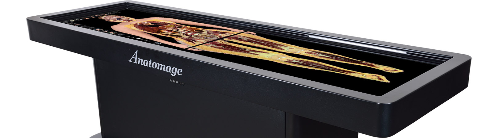
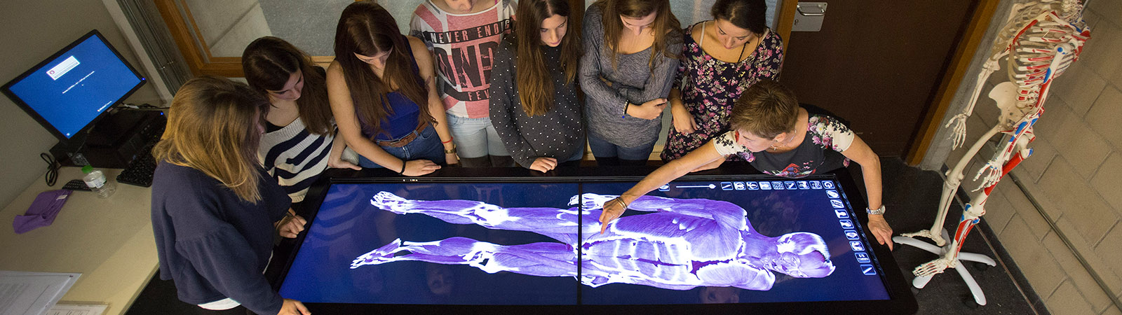
Still, the Table offers a completely unique and effective educational tool that can be incorporated into any curriculum. The faculty and students can engage the subject with a great degree of immersion.

The Table can be a foundation or a supplement to any gross anatomy curriculum and teaching method. The faculty and students will be equally impressed with the visual realism and details of this life-sized virtual dissection table. The Table allows students to visualize skeletal tissues, muscles, organs and soft tissue. These various tissues and views can be customized by virtually slicing, layering, and segmenting the anatomy. This adds a new dimension of depth to the education that the students will receive. Whether the students have access to cadaver based dissections or model based dissections, The Table offers primary and supplemental information. Unlike a cadaver, the imaging data can be altered extensively and instantly restored and reused many times. The students will gain additional understanding of the spacial relationships of anatomy, locations of hard to identify structures, and will be able to better comprehend the relationships of multiple biological systems in the human body. With flexible annotation tools, institutions can create innovative programs, quizzes, and study methods.

Research Institutes

The Table offers a whole host of new research potential. This could be research directly related to the use of this new technology for academic teaching methods, patient education, case reviews, hospital team work and other similar studies where this new method of interaction could be assessed. It could also be used as a center piece for different researchers to come together and interact with data, images, presentations, and other media all at the same time.

Corporations

The Table adds a tremendous amount of prestige to any conference room. Companies can use it to impress their guests, business partners, or to win corporate relationships within their industry. When placed in a trade-show, this will be the most dramatic attention grabbing tool, pulling customers in and increasing the time they stay at the booth and ask questions. Due to its advanced educational foundation, this provides the highest quality experience and delivers a trustworthy and effective presentation to doctors and clients.

Software

The unique anatomy study software is developed by Anatomage based on the renowned and successful Invivo5 imaging software technology. The software has unique rendering technology that allows for volume rendering, 3D mesh rendering and photographic rendering to simultaneously blend with each other in and accurate and consistent 3D space. When blended together it would be hard to distinguish which portion is volume rendering and which portion is surface rendering, thus creating a very advanced visualization that is unmatched.

Anatomage-virtual-dissection-table3


3D Digital Anatomy Library

Also available for expanded utilization of the Table is a complete library collection of 100 real-life clinical scans involving hospital emergency cases, pathology cases, and scans featuring routine medical examinations where CT imaging modality is the standard.


Custom Use

The Table is not limited to the provided data set. Users can open any radiology scan data for 3D visualization. The system is based on FDA approved medical imaging software, and it is compatible with any DICOM data, such as CT or MRI data. Users can zoom, rotate and slice with the fingers just like the anatomy data.

Client Rating: 4.8 based on 248 Clients